

BOARD-CERTIFIED PATHOLOGIST
AP/CP · Fellowship-trained · 14 yrs experience
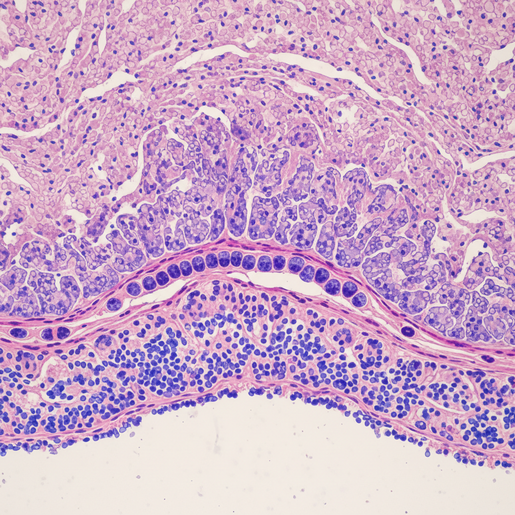
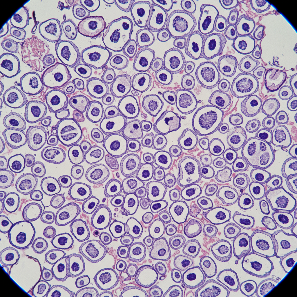
H&E · 40× magnification
Invasive ductal carcinoma, grade II
Your diagnosis, explained by the pathologist who read it.
Second opinions and primary reads. Board-certified. 48 hours.
What happens to your biopsy, step by step
Medicine usually hides this process. We don't. Here is exactly what happens on both sides of the microscope — from the moment your slides arrive to the moment you understand your results.
Submit your slides
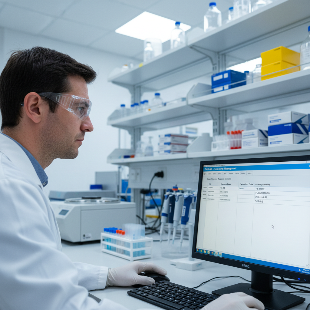
You or your physician ships the paraffin-embedded blocks or unstained slides to our secure receiving lab. A prepaid shipping kit arrives within 24 hours of request. Your portal account is created automatically — no forms, no fax.
Accession and prepare
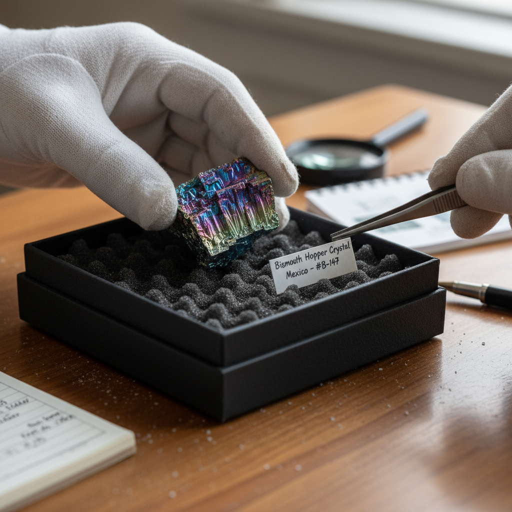
Upon receipt, our laboratory technician logs the specimen into our LIS, verifies chain of custody, and cuts additional sections if needed. Every slide gets a unique accession number tied to your case — nothing is anonymous to us.
You receive a notification
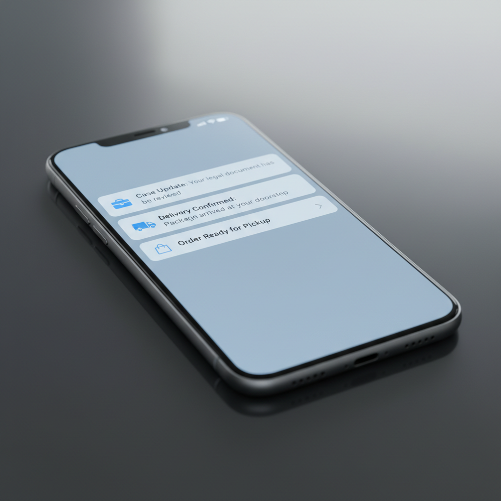
A text and email arrive: "Your slides are under the microscope." You can check your portal for real-time status — Received, Under Review, Report Drafted, Ready. No more calling the office and waiting on hold.
Examine under magnification
The assigned pathologist reviews the slides at 4×, 10×, 20×, and 40× magnification. Special stains — IHC, FISH, PAS — are ordered if the initial H&E raises questions. Every field of view is documented.
Join a video call
At the scheduled time, the pathologist who personally read your slides joins a secure video call. They share their screen — you see the actual tissue. They walk you through every finding in language calibrated to you, not a textbook.

Annotate and record
The pathologist annotates key regions of the slide — marking margins, grading differentiation, calling out any incidental findings. A screen-recorded consultation is added to your permanent record for your referring physician.
A plain-language report
Two documents land in your portal: the formal pathology report (for your physician) and a plain-language summary written for you. No jargon. No abbreviations without definitions. A glossary is included for every technical term used.

Dual-format diagnostic report
The formal report follows CAP-compliant synoptic formatting with ICD-10 coding, TNM staging where applicable, and specific recommendations for further workup. A signed PDF is transmitted directly to your referring physician's EHR.

One biopsy. Two ways of reading it.
SURGICAL PATHOLOGY REPORT
Accession: SW-2024-1187 · Received: 02/14/2026 09:42
Clinical History
58F. Right breast mass, 2.1 cm on ultrasound. Core needle biopsy, 4 cores. Prior mammogram: BI-RADS 4C.
Gross Description
Received in formalin: four tan-pink cylindrical cores ranging 1.2–1.6 cm in length, 0.1 cm in diameter. Entirely submitted in 2 cassettes.
Microscopic Description
Sections show infiltrating carcinoma with ductal differentiation. Tumor cells are arranged in nests and cords with moderate nuclear pleomorphism, prominent nucleoli, and frequent mitotic figures (11/10 HPF). Surrounding desmoplastic stroma. No lymphovascular invasion identified on H&E. ER: 85% strong positive. PR: 40% moderate positive. HER2: 1+ (negative). Ki-67: 28%.
Diagnosis
RIGHT BREAST, CORE NEEDLE BIOPSY: Invasive ductal carcinoma, NOS, Nottingham grade II (tubules 3, nuclei 2, mitoses 2; score 7/9). ER+/PR+/HER2−. Ki-67 28%. Recommend multidisciplinary tumor board review.
Dr. Margaret Osei-Bonsu, MD
AP/CP · Breast Pathology Fellowship · Signed 02/15/2026 16:08
What We Found, In Plain English
Written for you — no medical degree required
The short version
The biopsy found breast cancer. It is called "invasive ductal carcinoma" — the most common type of breast cancer. It was caught at a stage where treatment options are good.
What "grade II" means
Cancer cells are graded 1 to 3 based on how different they look from normal cells. Grade II is the middle — not the slowest-growing, not the fastest. Your pathologist scored three features of the cells and totaled them to reach this grade.
What the hormone tests mean
Your cancer tested positive for estrogen receptors (ER+) and progesterone receptors (PR+). This is good news — it means the cancer responds to hormone-blocking treatments, which are generally well-tolerated and effective.
What HER2-negative means
HER2 is a protein that can make some cancers grow faster. Your cancer does not overexpress HER2, which means a whole category of aggressive behavior is off the table.
What happens next
Your oncologist will present your case at a tumor board — a meeting of specialists who review cases together. They will use this report to design a treatment plan. You should ask about breast MRI, genetic testing (BRCA), and whether a lumpectomy or mastectomy is right for you.
"I had 14 minutes with my oncologist and left more confused than when I arrived. Dr. Osei-Bonsu spent 40 minutes on video with me and I finally understood what was in my body. That call changed everything."
— Patient, age 58, North Carolina
48 hrs
Average turnaround time
From specimen receipt to signed report
2,400+
Cases completed
Across 38 states since 2021
97%
Referring physician satisfaction
Based on post-consultation survey
100%
Board-certified pathologists
AP/CP, fellowship-trained specialists
From physicians who've referred, and patients who've been seen
I practice in a rural county without a local pathologist. Pathways has become my co-pilot for every ambiguous biopsy. The 48-hour turnaround means my patients don't spend a weekend in dread.
Dr. James Whitfield, MD
Family Medicine · Rural Health Clinic, Asheville, NC
Primary CareThe dual-format report is the differentiator. My patients come to their follow-up appointments having already read the plain-language summary. The conversation is completely different — they're partners, not passengers.
Dr. Priya Krishnamurthy, MD
Oncology · Mountain View Cancer Center, Flagstaff, AZ
OncologyOur critical access hospital couldn't justify a full-time pathologist. Pathways gives us board-certified reads on every case at a fraction of the cost, with a video consultation that my patients consistently describe as the most informative conversation of their cancer journey.
Dr. Robert Carvalho, MD
Chief of Medicine · Cascade Valley Hospital, WA
Hospital AdministrationInformation is medicine, too.
We've spent years watching people receive diagnoses without context. These guides exist to change that.
The Patient Guide to Biopsy Pathology
Plain-language explanation of what happens after a biopsy is sent for pathology review — written for patients, caregivers, and anyone who found this page at midnight.
Referring Provider Toolkit
CPT code reference, requisition forms, specimen shipping protocols, and EHR integration guide — everything your practice needs to start referring within 24 hours.
Both resources are free. No sales calls. No account required. For urgent cases, call our clinical intake line: 1-800-555-1847